
Skin Structure infographic LifeMap Discovery
Dermis. Definition. Fibrous and elastic tissue, provides strength and elasticity to the skin and supports the epidermis, home to hair follicles, glands, nerves etc. Location. Term. Papillary Layer. Definition. Upper dermal layer, provides the epidermis with nutrients and regulates body temperature. Location.

34 Label The Skin Diagram Labels 2021
Definition Middle layer of the skin; contains collagen; location of sweat & sebaceous glands and nerve endings and capilaries Location Term Epidermis Definition outermost layer of the skin;composed of squamous epithelium; contains keratin Location Term subcutaneous layer Definition
Dermis Layers, Papillary Layer, Function Epidermis
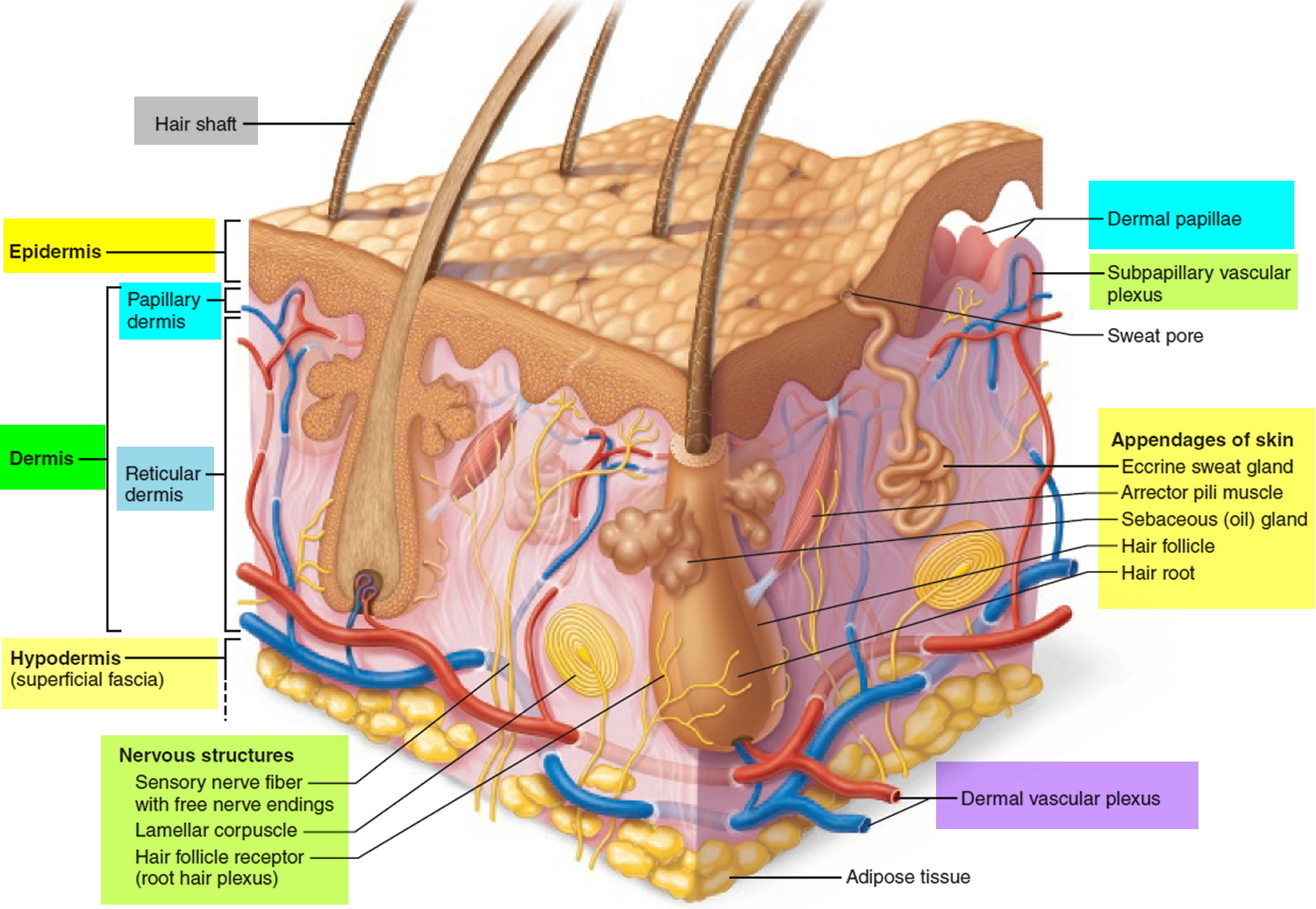
Figure 5.1.1 - Layers of Skin: The skin is composed of two main layers: the epidermis, made of closely packed epithelial cells, and the dermis, made of dense, irregular connective tissue that houses blood vessels, hair follicles, sweat glands, and other structures.

The structure of the skin is composed of two layers (1) the epidermis... Download Scientific
The Epidermis The epidermis is composed of keratinized, stratified squamous epithelium. It is made of four or five layers of epithelial cells, depending on its location in the body. It does not have any blood vessels within it (i.e., it is avascular). Skin that has four layers of cells is referred to as "thin skin."

Dermatology Diagram Show Human Skin Structure Stock Illustration Download Image Now Anatomy
Anatomy of the Skin Click Image to Enlarge Facts about the skin The skin is the body's largest organ. It covers the entire body. It serves as a protective shield against heat, light, injury, and infection. The skin also: Regulates body temperature Stores water and fat Is a sensory organ Prevents water loss Prevents entry of bacteria
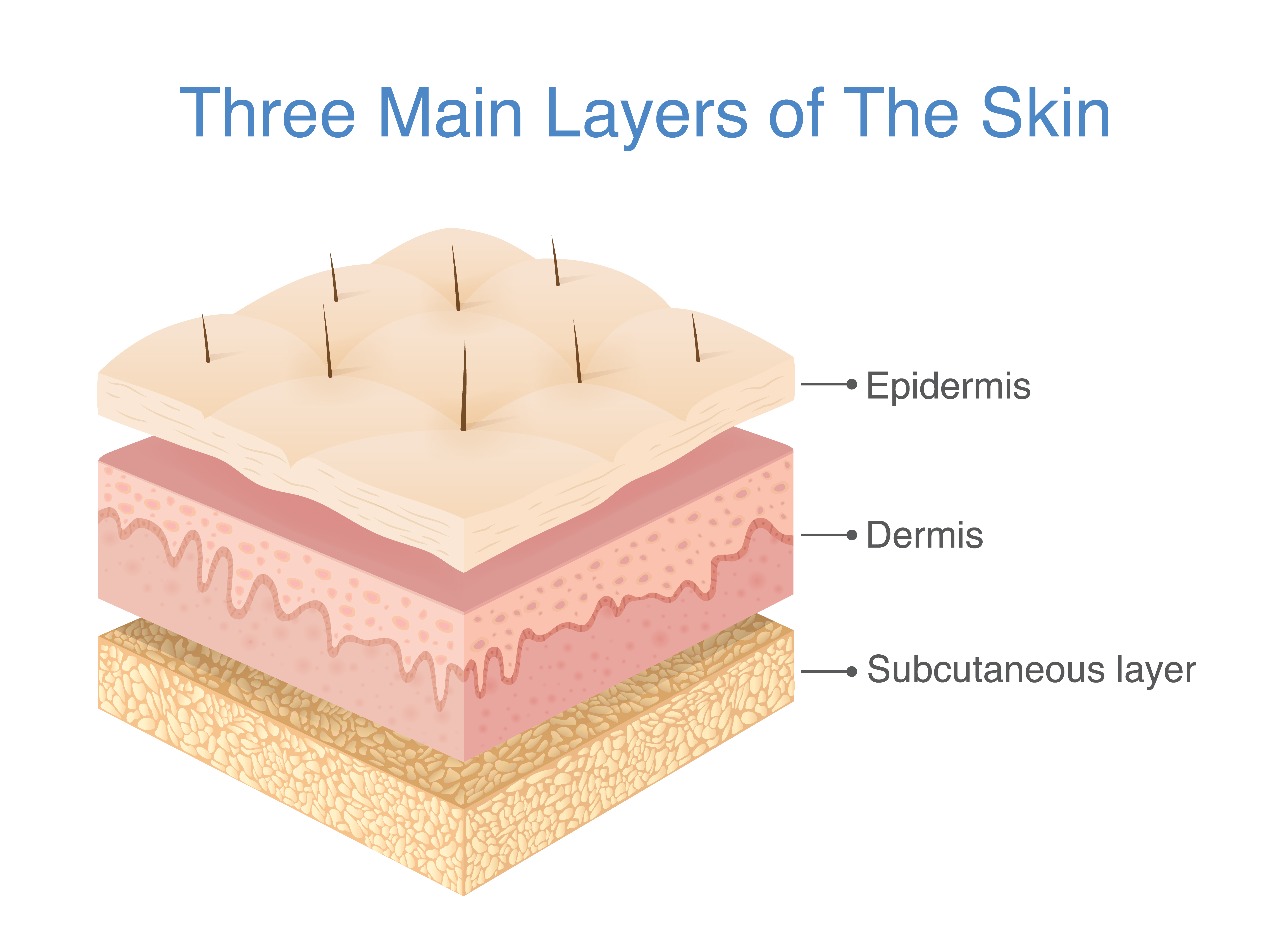
What Are The 3 Layers Of Skin? SkinMindBalance
'Skin Diagram || How to draw and label the parts of skin' is demonstrated in this video tutorial step by step.The sense of touch had received supreme importa.

Human skin diagram Subcutaneous tissue, Skin structure, Epidermis
Skin Labeling by marthamae 229,123 plays 17 questions ~40 sec English 17p 109 3.87 (you: not rated) Tries Unlimited [?] Last Played December 6, 2023 - 05:41 PM There is a printable worksheet available for download here so you can take the quiz with pen and paper. From the quiz author Epidermis, Dermis, Hypodermis Remaining 0 Correct 0 Wrong 0
Normal human skin [Biologie de la peau]
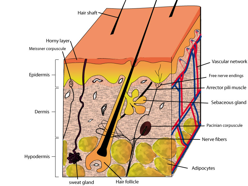
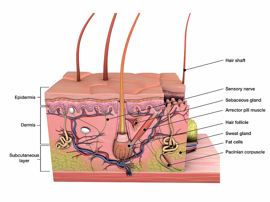
This article will describe the anatomy and histology of the skin. Undoubtedly, the skin is the largest organ in the human body; literally covering you from head to toe. The organ constitutes almost 8-20% of body mass and has a surface area of approximately 1.6 to 1.8 m2, in an adult. It is comprised of three major layers: epidermis, dermis and.
Anatomy Of Human Skin With Labels Photograph by Hank Grebe Pixels
The Epidermis The Dermis Hypodermis The number of skin layers that exists depends on how you count them. You have three main layers of skin—the epidermis , dermis, and hypodermis (subcutaneous tissue). Within these layers are additional layers. If you count the layers within the layers, the skin has eight or even 10 layers.

Structure Of Skin Skin Structure and Function LearnFatafat
Anatomy of the Skin Skin Facts about the skin The skin is the body's largest organ. It covers the entire body. It serves as a protective shield against heat, light, injury, and infection. The skin also: Regulates body temperature Stores water and fat Is a sensory organ Prevents water loss Prevents entry of bacteria
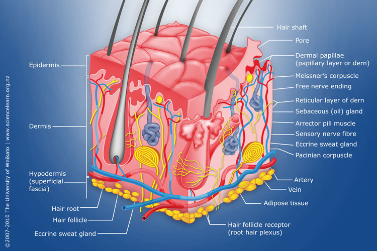
Diagram of human skin structure — Science Learning Hub
Study aids. Related quizzes:. Physiology of the skin, Quiz 1 - Now you know the parts of the skin, learn how they function.; The anatomy of bones, Quiz 1 - Learn the anatomy of a human long bone.; The anatomy of muscle, Quiz 1 - How much do you know about the anatomy of a the different muscle types?; Images and pdf's:. Just in case you get tired of looking at the screen we've provided images.
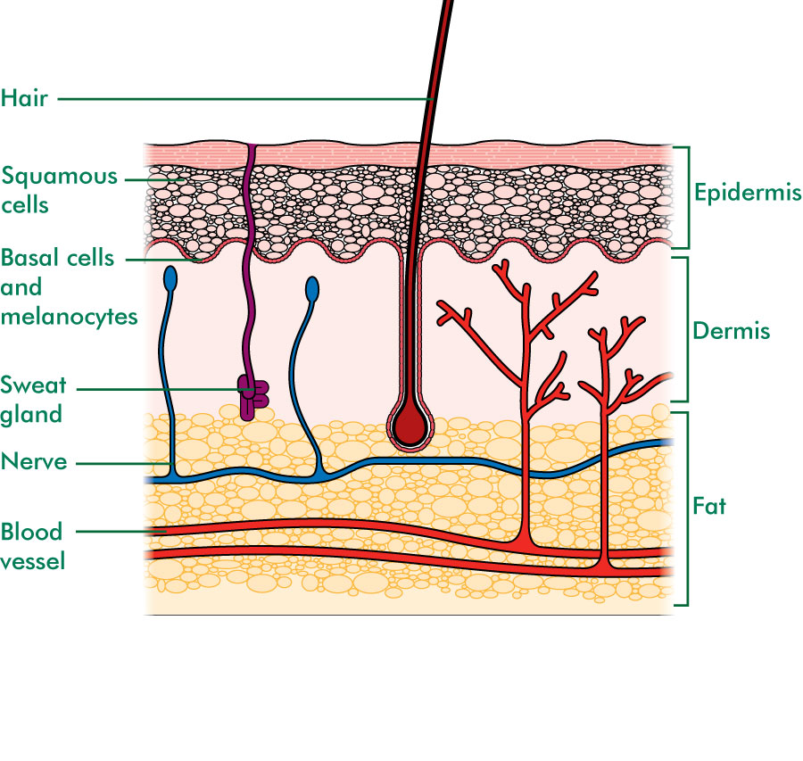
The skin Understanding cancer Macmillan Cancer Support
This online quiz is called label the skin diagram. It was created by member k8e1404 and has 12 questions.

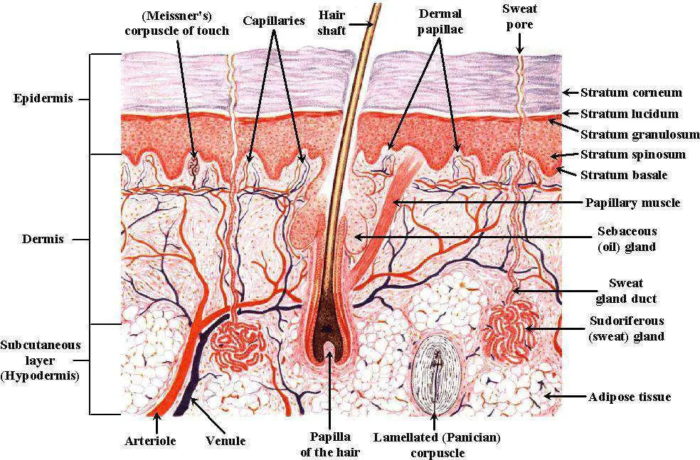
loadBinary_006.gif (992×779) Skin anatomy, Integumentary system, Human integumentary system
Figure 1. Layers of Skin. The skin is composed of two main layers: the epidermis, made of closely packed epithelial cells, and the dermis, made of dense, irregular connective tissue that houses blood vessels, hair follicles, sweat glands, and other structures.

Skin Definition, Structure And Functions Of Skin
Biology Important Diagrams Skin Diagram Skin Diagram The largest organ in the human body is the skin, covering a total area of about 1.8 square meters. The skin is tasked with protecting our body from external elements as well as microbes. Interesting Note:
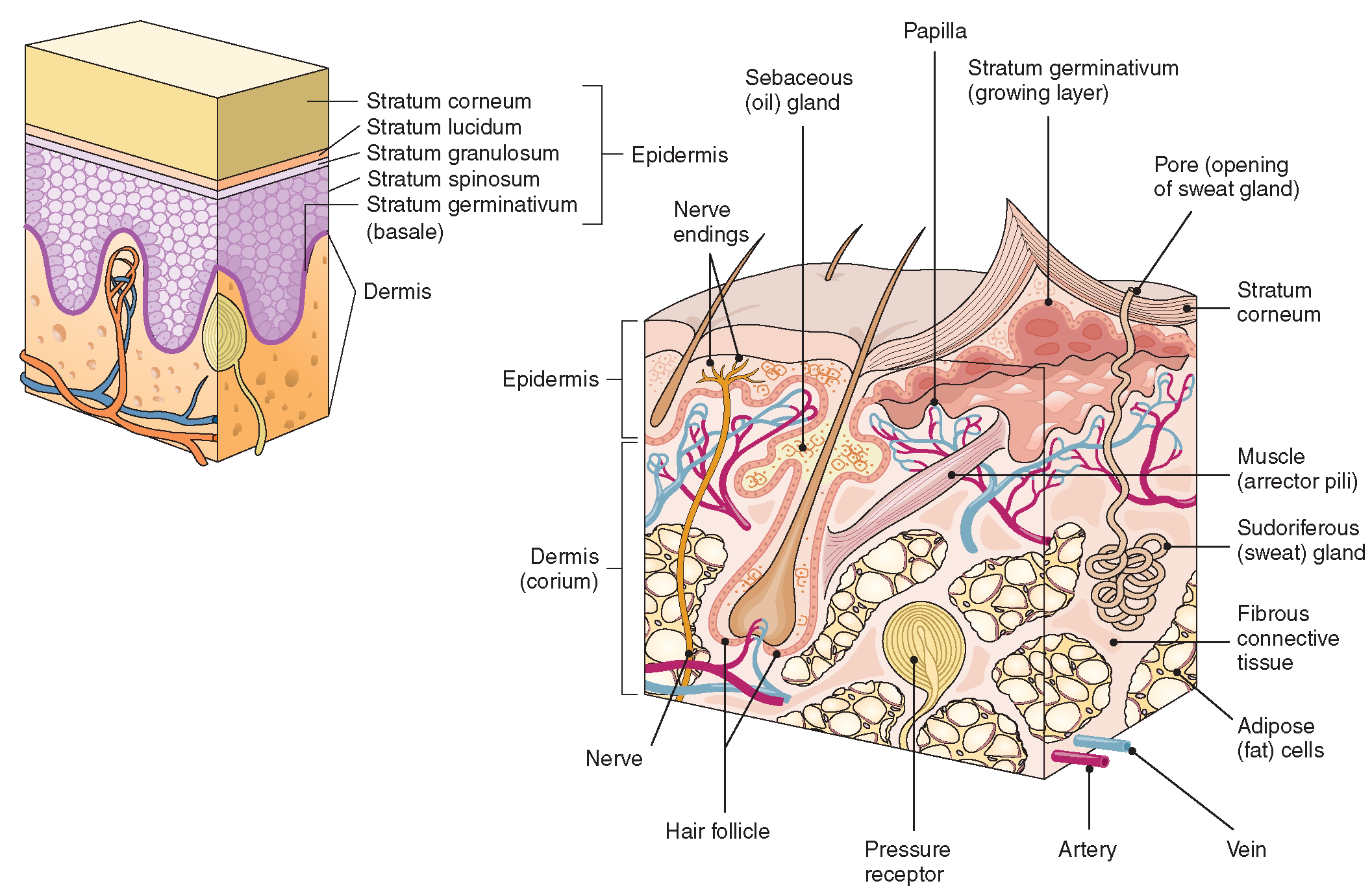
The Integumentary System (Structure and Function) (Nursing) Part 1
Skin diagram unlabeled Skin anatomy Specialized integumentary system quizzes Sources + Show all Integumentary system quiz and answers One of the best ways to start learning about a new system, organ or region is with a labeled diagram showing you all of the main structures found within it.
Skin diagram labeled
Functions. The skin has a significant capacity for renewal and crucial roles for the normal functioning of the human body. It is an effective barrier against potential pathogens and protects against mechanical, chemical, osmotic, thermal and ultraviolet radiation damage (through melanin). The skin also takes part in a variety of biochemical synthetic processes, such as vitamin D production.